

Why Are Neurons Necessary For Animal Systems, And Why Are They Only Found In Animal Systems?
Learning Objectives
- Describe dissimilar types of nervous systems in dissimilar creature lineages
- Identify and describe bones structures and functions of the key and peripheral nervous systems
- Describe the roles of and differentiate between the divisions of the vertebrate nervous arrangement (afferent, efferent, somatic, autonomic, sympathetic, parasympathetic)
- Define learning and memory, and explain the roles of the hippocampus, the cerebral cortex, neuroplasticity, and synaptic plasticity in learning and memory
Neurons, Nerves, and the Nervous System
The information below was adapted from OpenStax Biological science 35.0 and Khan Academy Overview of neuron structure and role. All Khan University content is available for gratis at www.khanacademy.org
When you're reading this website, your nervous organization is performing several functions simultaneously. The visual organization is processing what is seen on the page; the motor organization controls the click of the mouse; and (if you're lucky) the prefrontal cortex maintains your attention. Fifty-fifty primal functions, like breathing and regulation of trunk temperature, are controlled past the nervous system. A nervous system is an organism'south control center: it processes sensory information from outside (and inside) the body and controls all behaviors: from eating to sleeping to finding a mate.
The nervous system is made up of neurons, specialized cells that can receive and transmit chemical or electric signals, and glia, cells that provide support functions for the neurons by playing an information processing role that is complementary to neurons. Nerves are bundles of nervous tissue, often containing hundreds to thousands of axons wrapped in connective tissue. Nerves in the peripheral nervous system (PNS) carry data to and from neurons in the the primal nervous system (CNS), where data is integrated and candy.
In that location are three different classes of neurons that make up the nervous system:
- Afferent neurons (as well called sensory neurons) get information about what's going on within and outside of the torso and bring that information into the CNS so it tin can exist processed. For instance, if you picked upward a hot coal, sensory neurons with endings in your fingertips would convey the information to your CNS that it was really hot
- Efferent neurons (also called motor neurons) get information from other neurons and convey commands to your muscles, organs and glands. For instance, if you picked upwards a hot coal, it motor neurons innervating the muscles in your fingers would cause your hand to allow get.
- Interneuons, which are found just in the CNS, connect one neuron to another. They receive information from other neurons (either sensory neurons or interneurons) and transmit information to other neurons (either motor neurons or interneurons). For case, if yous picked up a hot coal, the bespeak from the sensory neurons in your fingertips would travel to interneurons in your spinal cord. Some of these interneurons would signal to the motor neurons decision-making your finger muscles (causing you to let become), while others would transmit the signal upward the spinal cord to neurons in the brain, where it would be perceived as pain. Interneurons are the most numerous class of neurons and are involved in processing information, both in simple reflex circuits (like those triggered by hot objects) and in more complex circuits in the brain. It would be combinations of interneurons in your brain that would allow y'all to draw the conclusion that things that looked similar hot coals weren't adept to option upwardly, and, hopefully, retain that information for future reference.
The material in the nervous system tin also exist classified based on whether it containswhite matter (myelinated axons) andgrey matter (uunmyelinated axons and prison cell bodies).
Diverseness of Nervous Systems
The data below was adapted from OpenStax Biology 35.ane
Nervous systems throughout the creature kingdom vary in construction and complexity, every bit illustrated by the variety of animals shown below:
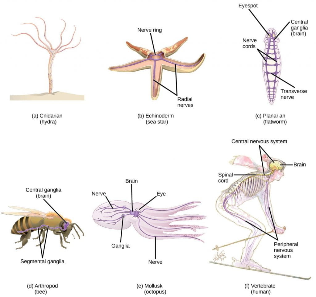
Nervous systems vary in structure and complexity. In (a) cnidarians, nerve cells form a decentralized nervus net. In (b) echinoderms, nervus cells are bundled into fibers called nerves. In animals exhibiting bilateral symmetry such as (c) planarians, neurons cluster into an anterior brain that processes information. In addition to a brain, (d) arthropods have clusters of nervus cell bodies, called peripheral ganglia, located forth the ventral nerve cord. Mollusks such as squid and (e) octopi, which must hunt to survive, have complex brains containing millions of neurons. In (f) vertebrates, the brain and spinal cord comprise the central nervous system, while neurons extending into the rest of the body contain the peripheral nervous system. (credit: OpenStax Biology, east: modification of work past Michael Vecchione, Clyde F.Due east. Roper, and Michael J. Sweeney, NOAA; credit f: modification of piece of work by NIH)
- All animals have a true nervous system except sea sponges.
- Cnidarians, such equally jellyfish, lack a truthful brain but accept a system of separate but connected neurons called a nervus net.
- Echinoderms, such equally sea stars, take neurons that are arranged into fibers called nerves.
- Flatworms of the phylum Platyhelminthes accept both a CNS made upwardly of a pocket-sized brain and two nerve cords, and PNS containing a system of fretfulness that extend throughout the body.
- The insect nervous organization is more complex but also adequately decentralized, with a brain, ventral nerve cord, and ganglia (clusters of connected neurons). These ganglia can control movements and behaviors without input from the brain.
- Cephalopods, such every bit octopi, may accept the most complicated of invertebrate nervous systems, with neurons that are organized in specialized lobes and eyes that are structurally similar to vertebrate species.
- Compared to invertebrates, vertebrate nervous systems are more complex, centralized, and specialized. While in that location is bang-up variety amidst different vertebrate nervous systems, they all share a bones construction: a CNS that contains a encephalon and spinal cord and a PNS made up of peripheral sensory and motor nerves.
One interesting deviation between the nervous systems of invertebrates and vertebrates is that the nervus cords of many invertebrates are located ventrally (along the belly) whereas the vertebrate spinal cords are located dorsally (along the back). There is debate amongst evolutionary biologists as to whether these different nervous system plans evolved separately or whether the original invertebrate body programme system somehow "flipped" during the evolution of vertebrates.
The Central Nervous Organisation in Vertebrates
The information below was adapted from OpenStax Biology 35.3 and Khan Academy Overview of neuron structure and function. All Khan Academy content is available for free at www.khanacademy.org
For the rest of this class session, we'll focus on the vertebrate nervous system. In vertebrates, the nervous system tin be broadly divided into two sections:
- a central nervous system (CNS) consisting of:
- the brain, a structure thatprocesses data, composed of inter-connected neurons and glial cells
- the spinal string, a structure that transmits information, consisting of a thick bundle of nervus tissue that carries information about the trunk to the brain and from the brain to the body
- a peripheral nervous system (PNS) that collects information and sends commands, containing fretfulness that extend to and from the spinal cord and are divided into:
- afferent nerves that collect sensory information from the body and transmit it to the CNS; afferent nerves are also sometimes chosen sensory nerves
- efferent nerves that carry commands from the CNS to the body; efferent nerves are also sometimes called motor nerves

The vertebrate nervous arrangement consists of a CNS and PNS. Image credit, https://www.khanacademy.org/scientific discipline/biology/ap-biological science/human-biology/neuron-nervous-system/a/overview-of-neuron-construction-and-role, modified from "Nervous organization diagram," by Medium69 (CC Past-SA four.0).
Anatomy of the Brain
The encephalon is the role of the central nervous system that is contained in the cranial cavity of the skull. It floats in a fluid called cerebrospinal fluid (CSF), which is filtered from arterial blood. CSF acts as a cushion and daze cushion, and makes the encephalon neutrally boyant. Ependymal cells (recollect those?) help broadcast the CSF to distribute and exchange chemic substances throughout the brain and into the spinal cord. The ependymal cells line the fluid-filled cavities in the eye of the brain, calledventricals, which too contains CSF.
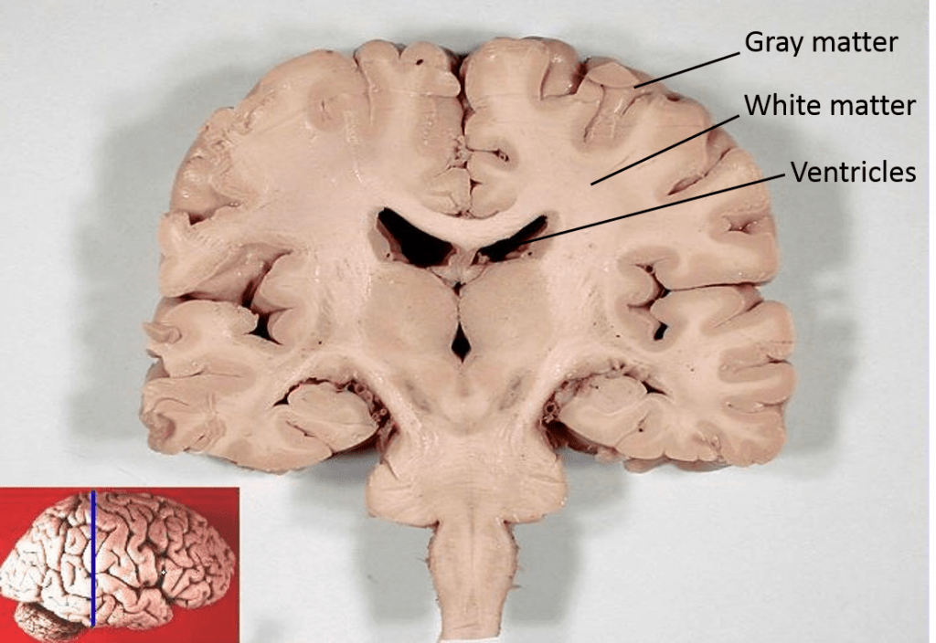
Modified from: John A Aggravate, PhDDep't. of Cellular Biology & Anatomy, Louisiana State University Wellness Sciences Center Shreveport – http://www.healcentral.org/healapp/showMetadata?metadataId=40566 (Internet Archive of file description page), CC By 2.five, https://commons.wikimedia.org/w/index.php?curid=879154
At the general anatomy level (shown above), the brain is organized with white matter (myelinated axons) toward the inside of the brain, and grayness matter (unmyelinated axons and jail cell bodies) on the outside of the brain. Gray matter represents the information-processing centers of the brain, and white thing represents the networking between these processing centers. These processing centers include (but are not express to!) the cerebrum and cerebral cortex, the diencephalon, the cerebellum, and the brain stem, further described below.
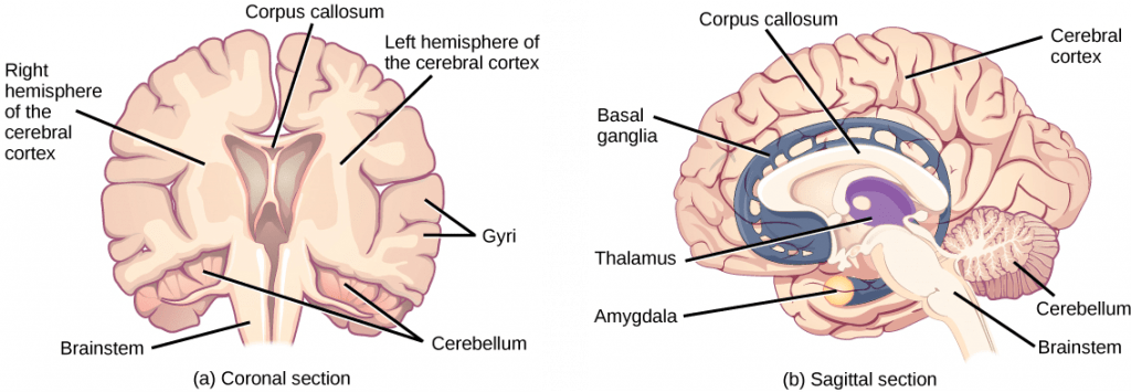
The vertebrate brain includes the cerebrum, cerebral cortex, diencephalon, cerebellum, and the encephalon stem. Prototype credit: OpenStax Biology.
- Thecerebrum and cerebral cortex make upwards the bulk of the human encephalon. The outermost part of the cerebrum is a thick slice of nervous system tissue chosen the cerebral cortex, which is folded into hills called gyri (singular: gyrus) and valleys chosen sulci (atypical: sulcus). The cortex is made upwardly of ii hemispheres (right and left) and iv lobes (frontal, parietal, temporal, occipital). The ii hemispheres are joined by a thick fiber bundle called the corpus callosum (Latin: "tough body") which connects the 2 hemispheres and allows information to be passed from one side to the other. Although there are some encephalon functions that are localized more to 1 hemisphere than the other, the functions of the 2 hemispheres are largely redundant.
- The diencephalon controls homeostasis and acts as a relay station, transmitting sensory information from from sensory neurons to the cerebrum. The structures located in the diencephalon that regulate these processes include the thalamus and the hypothalamus. The thalamus (Greek for "inner chamber") acts equally a gateway to and from the cortex. Information technology receives sensory and motor inputs from the body and also receives feedback from the cortex, and helps regulate consciousness, arousal, and sleep states. Beneath the thalamus is the hypothalamus, which controls the endocrine system by sending signals to the pituitary gland, a pea-sized endocrine gland that releases several unlike hormones that affect other glands likewise as other cells. This relationship ways that the hypothalamus regulates important behaviors that are controlled by these hormones. The hypothalamus is the body'southward "thermostat" (thinknegative feedback loop); information technology makes certain key functions like food and h2o intake, energy expenditure, and body temperature are kept at appropriate levels. Neurons within the hypothalamus also regulate circadian rhythms, sometimes chosen sleep cycles.
- The cerebellum (Latin for "little brain") sits at the base of the brain on superlative of the brainstem. The cerebellum controls balance and aids in coordinating circuitous movements and learning new motor tasks.
- The brainstem connects the rest of the brain with the spinal cord. Motor and sensory neurons extend through the brainstem assuasive for the relay of signals between the brain and spinal cord. The brainstem controls several important functions of the trunk including alertness, arousal, breathing, blood pressure, digestion, heart rate, swallowing, walking, and sensory and motor data integration.
Each cortical hemisphere of the cerebrumcontains regions chosen lobes that are involved in dissimilar functions, and include the frontal, parietal, temporal, andoccipital lobes:
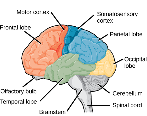
Sagittal, or side view of the human being encephalon shows the unlike lobes of the cerebral cortex. The frontal lobe is at the front center of the brain. The parietal lobe is at the top dorsum part of the brain. The occipital lobe is at the back of the brain, and the temporal lobe is at the bottom center of the brain. The motor cortex is the back of the frontal lobe, and the olfactory seedling is the bottom part. The somatosensory cortex is the front office of the parietal lobe. The brainstem is beneath the temporal lobe, and the cerebellum is beneath the occipital lobe. Image credit: OpenStax Biological science
The human cerebral cortex can be further subdivided into four lobes, each of which specializes in dissimilar functions:
- The frontal lobe is located at the forepart of the encephalon, over the eyes. This lobe contains the olfactory bulb, which processes smells. The frontal lobe also contains the motor cortex, which is important for planning and implementing move. Neurons in the frontal lobe likewise control cognitive functions like maintaining attending, speech, and controlling, and parts of this expanse are involved in personality, socialization, and assessing risk.
- The parietal lobe is located at the pinnacle of the brain. Neurons in the parietal lobe are involved in speech and too reading. Two of the parietal lobe'southward primary functions are processing somatosensation (impact sensations similar pressure, pain, rut, common cold) and processing proprioception (the sense of how parts of the trunk are oriented in infinite).
- The occipital lobe is located at the back of the encephalon. Information technology is primarily involved in vision: seeing, recognizing, and identifying the visual world. Sensory neurons transmit information from the optics at the forepart of the skull to the occipital lobe at the dorsum of the brain via the occipital nervus.
- The temporal lobe is located at the base of the brain past your ears and is primarily involved in processing and interpreting sounds. It likewise contains the hippocampus (Greek for "seahorse"); a structure that processes retentivity formation and is critical for learning.
Compared to other vertebrates, mammals take exceptionally large brains for their body size. An entire alligator'southward encephalon, for case, would fill virtually one and a one-half teaspoons. This increase in encephalon to body size ratio is particularly pronounced in apes, whales, and dolphins. While this increment in overall brain size doubtlessly played a role in the development of complex behaviors unique to mammals, it does not tell the whole story. Scientists have found a relationship betwixt the relatively high surface expanse of the cortex and the intelligence and complex social behaviors exhibited past some mammals. This increased surface surface area is due, in part, to increased folding of the cortical sheet (more sulci and gyri). For example, a rat cortex is very smooth with very few sulci and gyri. True cat and sheep cortices have more sulci and gyri. Chimps, humans, and dolphins have even more.
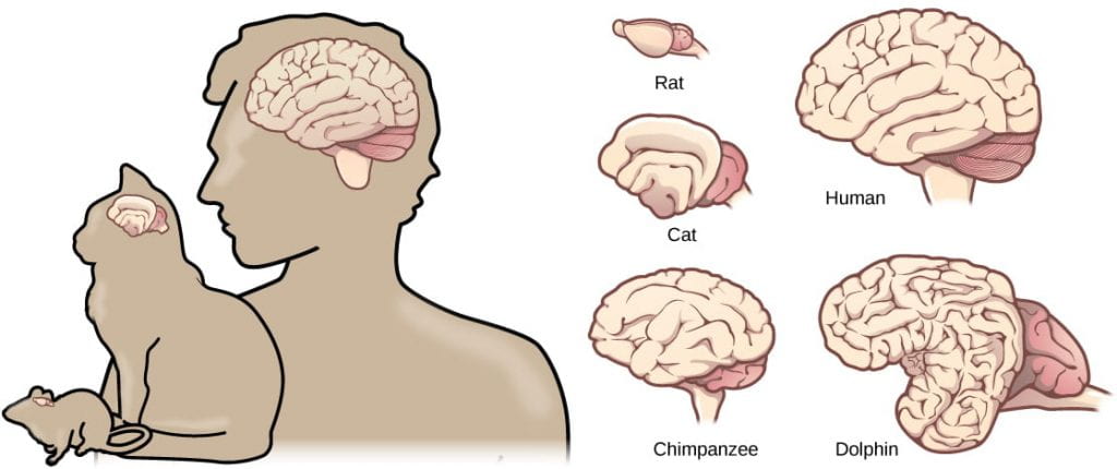
Mammals accept larger brain-to-body ratios than other vertebrates. Within mammals, increased cortical folding and surface area is correlated with complex beliefs. Image credit: OpenStax Biological science.
This video provides a summary of the functions of different parts of the vertebrate encephalon (lookout man the first 4 minutes):
Learning and Memory
The information below was adjusted from OpenStax Biology 35.2
One of the central functions that the brain performs is the process of learning and memory. Learning is the power to acquire new knowledge, and retentiveness is the ability to call up information technology afterward. Learning and retentivity involve both specific encephalon structures besides as sure neuronal processes. The current hypothesis states that specific neurons in the cerebral cortex are responsible for physically storing memories, and that learning and retentivity are mediated by both chemic and structural changes in the synapses of these neurons.
Brusk-term memories are thought to exist stored in the prefrontal cortex (office of the frontal lobe). Thehippocampus in the temporal lobe is essential for consolidating these short-term memories into long-term memories, just the memories are non really stored in the hippocampus. The precise location of retentivity storage is unknown, but it is though that different components of memories may be stored in dissimilar locations within the cerebral cortex, and that retrieval of long-term memories may involved the prefrontal cortex.
Storage and access are only half of the story for learning and retentivity; the other one-half is chemic and structural changes in synapses, or neural plasticity: germination of new and loss of existing neural connections. By the finish of embryogenesis in humans, half of all embryonic neurons undergo programmed cell death, and half of the initial synapses are lost. This bones neural architecture is then continually remodeled during the individual'south lifetime. How does neural plasticity relate to learning and memory? Chemical and structural changes in synapses (synaptic plasticity, synaptic pruning,synaptogenesis) mediate access to and forcefulness of these memories every bit follows:
- Neurogenesis , or the growth of new neurons. At 1 time, scientists believed that people were born with all the neurons they would always have, simply inquiry performed during the terminal few decades indicates that neurogenesis, the birth of new neurons, continues into machismo. Neurogenesis was first discovered in songbirds that produce new neurons while learning songs. For mammals, new neurons also play an of import office in learning: about k new neurons develop in the hippocampus (a encephalon construction involved in learning and memory) each day. While most of the new neurons will die, researchers found that an increment in the number of surviving new neurons in the hippocampus correlated with how well rats learned a new task. Interestingly, both do and some antidepressant medications also promote neurogenesis in the hippocampus. Stress has the contrary effect.
- Synaptogenesis, or the growth of new synapses between two existing neurons, andsynaptic pruning, or the destruction of existing synapses betwixt two neurons.
- Synaptic plasticity, or the strengthening or weakening of existing synaptic connections. Two processes in particular, long-term potentiation (LTP) and long-term low (LTD) are important forms of synaptic plasticity that occur in synapses in the hippocampus, a brain region that is involved in storing memories.
- Long-term potentiation (LTP) is the long-term strengthening of a synaptic connection. LTP is based on the thought that "cells that fire together wire together." At that place are diverse mechanisms underlying synaptic strengthening seen with LTP, including an increase in the amount of neurotransmitter released by the presynaptic neuron, and an increased response to the aforementioned amount of neurotransmitter past the postsynaptic neuron. LTP tin can upshot insensitization, where there is an increased response to the same external stimulus.
- Long-term depression (LTD) is essentially the contrary of LTP: it is a long-term weakening of a synaptic connectedness. While it may seem counterintuitive, LTD may be just every bit of import for learning and memory equally LTP: the weakening unused synapses allows for unimportant connections to be lost and makes the synapses that have undergone LTP that much stronger by comparison. LTD can result inhabituation, where at that place is a decreased response to the aforementioned external stimulus.
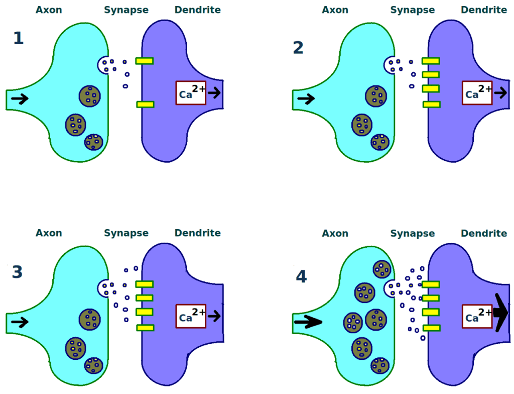
Long term potentiation can occur after repeated stimulation at a synaptic final (console ane) via several mechanisms, including production of more neurotransmitter receptors on the postsynaptic neuron (console 2) and production of more than neurotransmitter molecules by the presynaptic neuron (panel iii). A stronger connection between the neurons (panel 4) will occur as a result of either of these changes. Image credit: modification of work by Tomwsulcer – Own piece of work, CC0, https://commons.wikimedia.org/w/index.php?curid=15509518
This video provides a simplified overview of learning and memory in a usually used model organism for studying these processes:
And finally, this video provides a succinct overview of two of the mutual results of learning, sensitization or habituation:
The Spinal String
The data beneath was adjusted from OpenStax Biology 35.3 and Khan Academy Overview of neuron structure and office. All Khan Academy content is available for free at www.khanacademy.org
In order to learn from and answer to information, the encephalon must receive that data from the environment. Detection of information occurs via the PNS, and is relayed to the brain via the spinal cord (which is role of the CNS). The spinal cord connects to the brainstem and extends downwards the body through the spinal column. The spinal cord is a thick bundle of nerve tissue that carries data about the body to the brain and from the brain to the body. The spinal string is independent within the bones of the vertebrate column only is able to communicate signals to and from the body through its connections with spinal nerves (part of the peripheral nervous system).
A cross-department of the spinal cord looks similar a white oval containing a gray butterfly-shape. Myelinated axons make upward the white matter and neuron and glial cell bodies make upward the gray thing. Greyness matter is also composed of interneurons, which connect two neurons each located in different parts of the body. Relay of data to and from the spinal column is directional:
- Axons and cell bodies in the dorsal (facing the dorsum of the animal) spinal string convey mostly sensory information from the torso to the spinal string and then encephalon.
- Axons and cell bodies in the ventral (facing the front of the animal) spinal cord primarily transmit signals controlling movement from the brain to out to the torso.
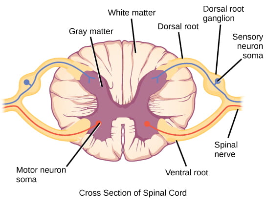
Spinal fretfulness comprise both sensory and motor axons. The somas of sensory neurons are located in dorsal root ganglia. The somas of motor neurons are found in the ventral portion of the gray matter of the spinal cord. Prototype credit: OpenStax Biological science
As noted previously, a nerve is not the same thing as a neuron; nerves are actually bundles of nervous tissue and tin contain hundreds to thousands of axons. Nerves contain both neuronal cells (axons and associated Schwann cells) and connective tissue (blood vessels and multiple layers of membranes bundling different groups of axons). The diagram below illustrates the general anatomy of a nerve:

Fretfulness contain blood vessels, neuronal tissues, and membranes composed of connective tissues bundling axons into different groups. Past Ranjit530 – Fatigued, CC BY-SA 3.0, https://commons.wikimedia.org/w/index.php?curid=29397234
The Peripheral Nervous System in Vertebrates
The information below was adapted from OpenStax Biological science 35.4
The spinal column transmits information to the CNS for processing. How does the information get to the spinal column to begin with? And how practise commands from the CNS go carried out? That is the task of nerves and the peripheral nervous system (PNS). If the CNS is similar the power plant of the nervous arrangement, creates the signals that control the functions of the body, and then the PNS is similar the wires that go to individual houses. Without those "wires," the signals produced by the CNS could not control the body (and the CNS would not be able to receive sensory information from the torso either).
The PNS is equanimous of the cranial and spinal fretfulness, and can be divided into the different divisions based on structure and role every bit follow:
- Theafferent (sensory) partition : collects incoming sensory data; made upward of cranial and spinal fretfulness that contain sensory neurons. Sensory neurons transmit sensory information from the skin, skeletal muscle, and sensory organs to the CNS. Without its afferent nervous system, an animal would exist unable to receive or process any data about its environment (what it sees, feels, hears, and so on).
- The efferent(motor) sectionalization: carries outgoing commands;made upwardly of cranial and spinal fretfulness that incorporate motor neurons. Motor neurons transmit letters near desired motion from the CNS to the muscles to make them contract. Motor neurons may exist under conscious or unconscious control. The efferent sectionalisation can exist further divided into:
- The somatic (conscious control) nervous system: sends motor commands from the CNS to voluntarily-controlled muscles; fabricated up of cranial and spinal nerves that contain motor neurons under conscious control. Without the somatic nervous organization, an beast would be unable to answer to its environment via controlled motor movements. Motor neurons of the somatic nervous system synapse with muscles under voluntary control, such as limb muscles.
- The autonomic (unconscious control) nervous system, which controls actual functions without conscious control; made of cranial and spinal nerves that contain motor neurons under unconscious control, such every bit the heart, smoothen musculus, and exocrine and endocrine glands. Can be further divided into two systems with opposing effects:
- The sympathetic nervous system, which controls the "fight or flying" reactions associated with the brusque-term stress response. Examples of functions controlled past the sympathetic nervous system include an accelerated heart rate and inhibited digestion. These functions help gear up an organism's trunk for the concrete strain required to escape a potentially dangerous situation or to fend off a predator. The sympathetic nervous organization alters the behavior of may organs directly via synapsed neurons, including the adrenal glands which then release norepinephrine and epinephrine into the blood stream. These hormones and then cause further changes, including dilation of the trachea and bronchi (making it easier for the animal to breathe), increasing heart charge per unit, and moving claret from the pare to the eye, muscles, and encephalon (so the animal can think and run).
- The parasympathetic nervous organization:controls the "balance and digest" activities involved in conserving and restoring energy. The parasympathetic nervous organisation resets organ office subsequently the sympathetic nervous system is activated (the common adrenaline dump you lot experience after a "fight-or-flight" event). Effects of the parasympathetic nervous organisation on target organs include slowing of heart rate, lowered blood force per unit area, and stimulation of digestion.
The opposing effects of the sympathetic vs parasympathetic nervous systems are illustrated below:
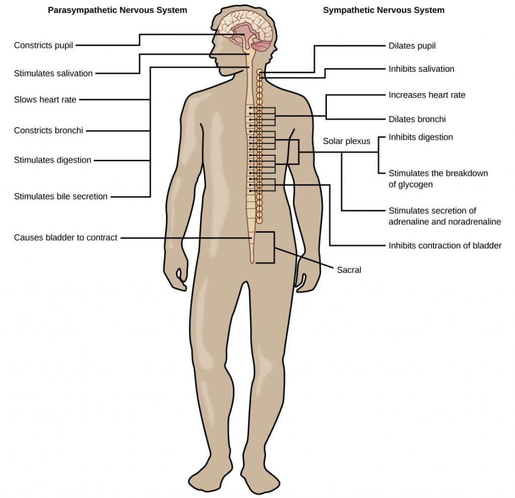
The sympathetic and parasympathetic nervous systems ofttimes have opposing effects on target organs, where the sympathetic system promotes the "fight or flying" response and the parasympathetic system promotes the "rest and assimilate" response. Prototype credit: OpenStax Biology
This video provides an overview of the CNS and PNS:
Reflex Arcs
Reflex arcs are an interesting phenomenon for because how the PNS and CNS work together. Reflexes are quick, unconscious movements, like automatically removing a hand from a hot object. Reflexes are so fast because they involve local synaptic connections in the spinal cord, rather than relay of information to the brain. For case, the knee reflex that a doctor tests during a routine concrete is controlled by a single synapse between a sensory neuron and a motor neuron. While a reflex may only require the interest of one or two synapses, synapses with interneurons in the spinal column transmit information to the brain to convey what happened after the event is already over (the knee jerked, or the manus was hot). So this means that the encephalon is not involved at all in themovement associated with the reflex, but it is certainly involved inlearningfrom the experience – nearly people just accept to impact a hot stove once to learn that they should never do information technology again!
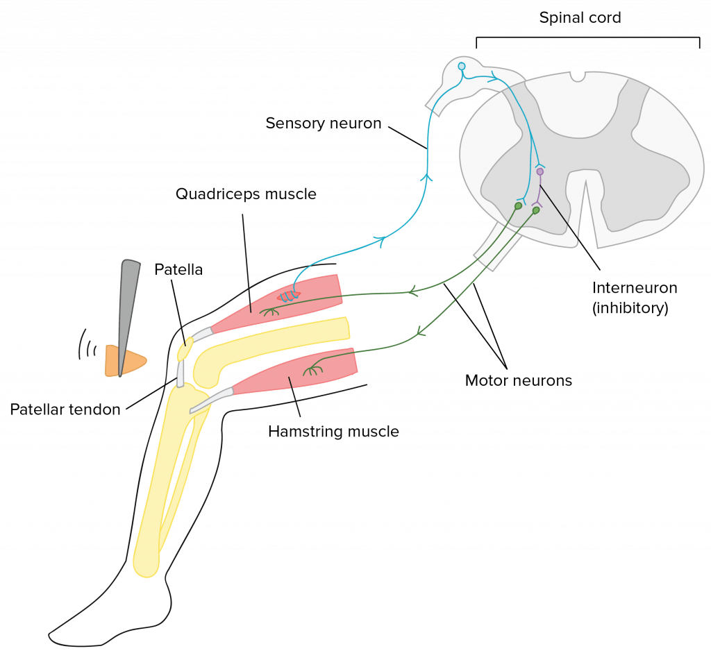
The simplest neuronal circuits are those that underlie muscle stretch responses, such as the genu-jerk reflex that occurs when someone hits the tendon below your knee joint (the patellar tendon) with a hammer. Tapping on that tendon stretches the quadriceps muscle of the thigh, stimulating the sensory neurons that innervate it to burn down. Axons from these sensory neurons extend to the spinal string, where they connect to the motor neurons that establish connections with (innervate) the quadriceps. The sensory neurons send an excitatory indicate to the motor neurons, causing them to burn down besides. The motor neurons, in turn, stimulate the quadriceps to contract, straightening the articulatio genus. In the knee-jerk reflex, the sensory neurons from a particular musculus connect directly to the motor neurons that innervate that aforementioned muscle, causing it to contract afterward it has been stretched. Epitome credit: https://www.khanacademy.org/science/biological science/ap-biological science/human-biology/neuron-nervous-arrangement/a/overview-of-neuron-construction-and-function, modified from "Patellar tendon reflex arc," past Amiya Sarkar (CC BY-SA four.0). The modified image is licensed nether a CC BY-SA four.0 license.
This video provides an overview of how reflex arcs work:
Source: https://organismalbio.biosci.gatech.edu/chemical-and-electrical-signals/nervous-systems/
Posted by: hardertraturness.blogspot.com
0 Response to "Why Are Neurons Necessary For Animal Systems, And Why Are They Only Found In Animal Systems?"
Post a Comment